CT Scan
(Cone beam computer tomography)
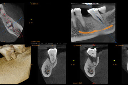
The Cone beam CT scan provides 3D images of the jaw structures and teeth with extraordinary accuracy and detail. It has become the gold standard for patient care in implant dentistry.The 3D images enable us to plan surgery so we are able to avoid causing damages to nerves and to vital structures, including blood vessels and the sinuses.
CAD/CAM Dentistry
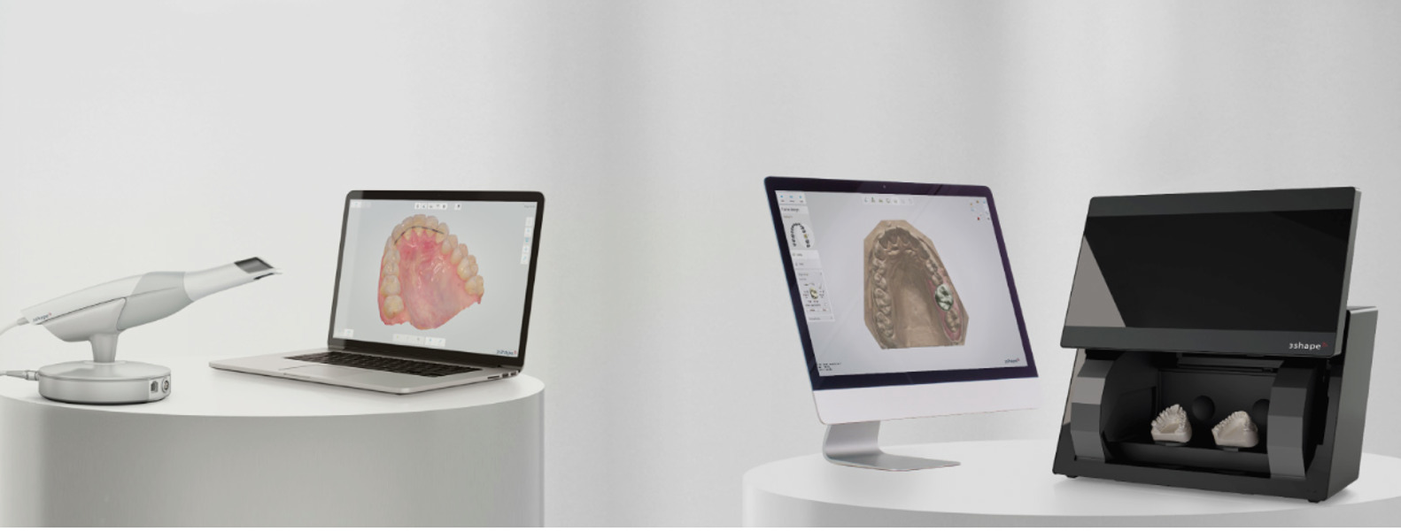
This technology and metal-free materials are used by dentists and dental laboratories to provide patients with milled ceramic crowns, veneers, onlays, inlays and bridges.
Today’s CAD/CAM restorations are better-fitting, more durable and more natural looking (multi-colored and translucent, similar to natural teeth) than previously machined restorations.
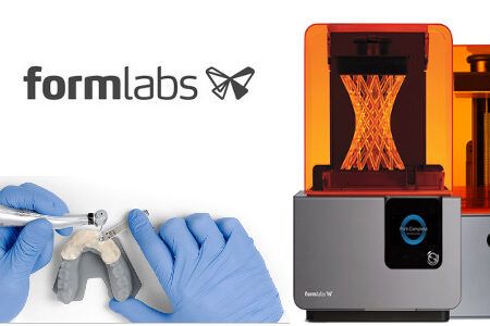
End-to-End Digital Dentistry:
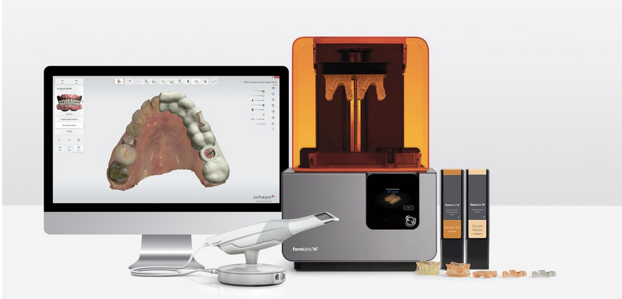
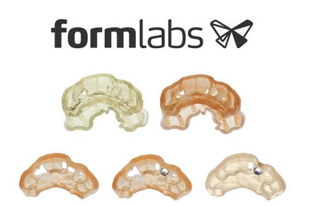
This new technology 3D printing allows you completely digitize your process; you are free to imagine new ways to advance care. Our solutions integrate with leading intraoral scanners and software to ensure predictable and repeatable results that provide perfect fit and occlusion.
Digital Radiography Advantage
Digital radiography typically reduces radiation exposure by 75% or more. That means your patients will notice a higher level of care when you use digital X-ray equipment. These features improve your ability to detect disease and it’s current state. They also provide immediate visuals for faster diagnosis.
The NOMAD PRO™ Wireless handheld dental X-ray systems.
The most advanced systems on the market today. The NOMAD Pro offers an enhanced user interface, preset and programmable exposure settings, and additional time-saving features. Its cordless design improves dental radiography speed and efficiency.
Proven safe, studies show yearly operator exposure is equivalent to or less than exposure from a typical wall-mounted X-ray.
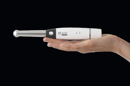
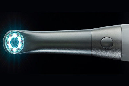
CS 1500 Intraoral Camera
The ideal communication tool delivering precise, true-to-life images with each shot, camera provides the visual evidence you need to educate patients and make more accurate diagnoses. . The camera delivers consistently clear, high-resolution dental digital photography images that can be easily shared with patients—so they see what you see and are more likely to understand treatment recommendations.
Platelet-rich fibrin (L-PRF)
Platelet-rich fibrin (L-PRF) is an autogenous matrix derived from the concentration of the patient’s blood platelets. A simplified chairside procedure results in the production of a fibrin membrane that is capable of stimulating the release of many important growth factors involved during wound healing processes that take place after surgery.
Platelet-rich fibrin (L-PRF) accelerates the body’s own normal healing processes making it an excellent choice for grafting applications , tissue graft , tooth extraction and implant.
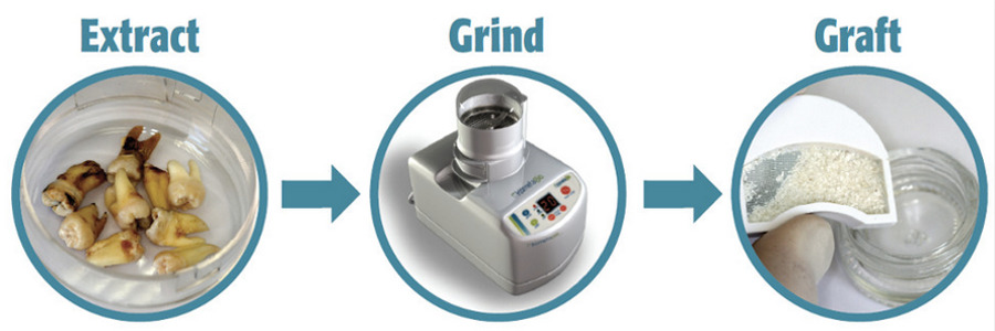
A revolutionary protocol to produce autologous dentin graft from a patient’s own tooth.
“We leveraged concepts of autologous grafting to optimize and simplify their use in dentistry. This resulted in dentists finally able to stop discarding extracted teeth and instead using them as Gold Standard bone graft.”.
The principles that guide us:
- Bone sets the tone for function and esthetics.
- Bone maintenance and repair is critical – graft whenever possible.
- Autologous grafting is the GOLD standard.
- Dentin is a proven and effective graft, 98% compatible with cortical bone.
- Every extraction site creates bone trauma and deficiency that must be corrected.
- It’s a clinical failure to discard an extracted tooth when it can be reused as a superior source for autologous graft.
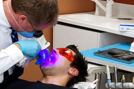
Velascope
A recent study involved oral cancer screenings for 85 male and female patients considered to be at risk for oral cancer. All patients were examined in two ways: a conventional clinical examination, consisting of palpation of the face and neck and an unassisted visual inspection of the oral cavity; and an examination of the oral.

